EyeWire’s First Scientific Discovery and Nature Paper
Today we are excited to share that EyeWire’s first scientific discovery has been published in Nature!
Why does it matter?
This discovery would not have been possible without you! Together, citizen scientists and gamers have made a significant contribution to neuroscience: we’ve uncovered one of the mechanisms that underlies a mammal’s ability to tell that something it sees is moving in a specific direction. Neuroscientists call this direction selectivity. Although this property sounds simple, its mechanism has been unclear since direction selectivity was first discovered more than 50 years ago.
Read the full paper here or check out this link to access the list of EyeWire player coauthors.
What have we done?
First, let’s survey your contribution. The EyeWire community has grown to 120K players from nearly 150 countries. In total, EyeWirers have reconstructed more than 100 neurons so far. Among them, 29 “Off”-type of starburst neurons were used in this paper (off-type means these cells react to dark stimuli). They were reconstructed by the elite players who passed the Starburst Challenge. The rest of the cells will be used in future projects.

Starbursts
“Starburst” is short for the full name “starburst amacrine cell”. It is one of many types of amacrine cells. There are other classes of cells, too. Photoreceptors, for example, are the cells that turn light into neuro-signals. Bipolar cells receive inputs from photoreceptors and in turn give out signals to the amacrine cells and ganglion cells. (“Bipolar” refers to the fact that the branches of these cells point in two opposite directions, or “poles”. It bears no relation to bipolar disorder). Amacrine cells make the neuronal network even more complex by connecting different bipolar cells, ganglion cells and even the amacrine cells themselves. Finally, the ganglion cells are output cells of the retina. They receive inputs from bipolar cells and amacrine cells and give outputs through their axons, which form the optic nerve and finally go into the brain. But this is just a rough description. Each class mentioned above has many types, and we know almost nothing of the connections among them. We had little idea how many bipolar partners starbursts have until this research.
Bearing this structure in mind, let’s consider what happens in the retina and how the signal reaches the starbursts when an animal sees things. When an object (seen as differentials in light) is on a spot of the animal’s field of view, the photoreceptors at the corresponding location are activated. Underneath each photoreceptor, there are a few bipolar cells, possibly in various types, connected to and receiving signal from the photoreceptors. In turn, the bipolar cells are connected to the starburst underneath them and send signals to it. Different types of bipolar cells send signals in different ways. We’ll get to why the different ways of different types matter in a moment.

First, two notes about the known function of starbursts:
1. Starbursts are in the amacrine family – and “amacrine” means “without axon” in Greek. They are different from textbook neurons. They have no axon. There’s no distinction between input and output branches and signals can be received and sent in one branch. Moreover it has different working mechanism. Textbook neurons propagate signals when the sum of inputs is large enough at a given moment. And the output is either on or off; it is like a digital signal. The amacrine cells and of course starbursts, on the other hand, propagate analog-like signals; their output can be strong or weak. The strength of output depends on the strength of sum of inputs. Combining, a starburst branch sends out signal onward according to the amount of input signals it gets.
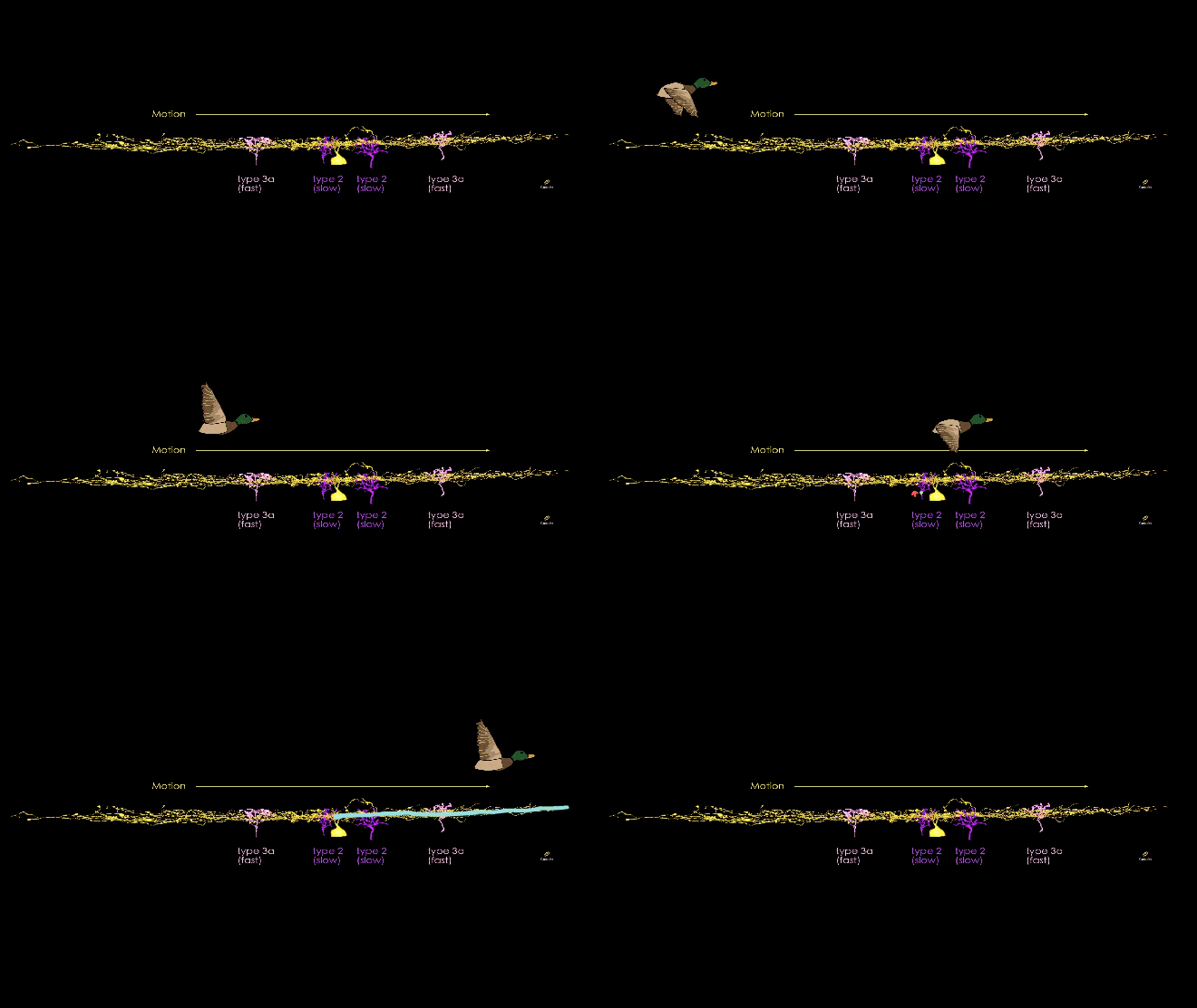
2. And such individual starburst branch working as an independent unit is direction selective. A starburst branch propagates a stronger signal downstream when a visual stimulus is moving along it in the direction away from cell body than when a stimulus is moving towards it, as illustrated by Figure 3 below. Say, for example, you are a mouse looking up at the sky, contemplating your cheese filled universe. Suddenly you see a duck fly off. The duck’s motion is represented as a time series of stimuli moving across your retina. Above, we saw what happens when a stimulus is on a spot of retina. Now the stimulus gradually moves on the retina. A queue of photoreceptors are activated in sequence, and propagate signals down to bipolars and then starbursts. That is, a starburst branch receive signals initiated by a queue of photoreceptors in order. But only as the order is in the direction moving away from the cell body to the tip does the starburst branch send out strong signal to its post-synaptic (downstream) neurons.
How is it possible? How does the sequential activation of bipolar cells turn into a stronger signal of the starburst? Here’s where the different types of bipolar cells come in.


Completing the Circuit
We proposed an answer to this question by analyzing the reconstructed cells from EyeWire. We analyzed 80 Off starbursts (29 of which were mapped by EyeWirers) and many Off bipolars that have potentials to make connections to the starbursts. Off bipolars come in at least five types (Figure 5). From the analysis, we found that bipolar cells of different types form synapses at different places on starburst cells.

Type 2, for instance, tend to synapse close to the starburst cell body. Type 3a, on the other hand, form synapses farther out on the branches as in Figure 6 and 7. Other types make fewer synapses than these two. It is an amazing discovery that of all the bipolars that have similar connections to the starbursts. Even more interesting is the two types’ having their own taste for pairing locations. So how does this result in direction selectivity?


While EyeWire community was mapping all these cells, a research group found a very cool thing about bipolar cells very recently. They found that Type 2 bipolars have this nifty time delay feature. A signal takes longer to cross through them than, say, Type 3a bipolar cells. Let’s suppose a signal enters Type 2 bipolar and another does Type 3a at the same time. But the output signals from the two don’t come out together because of the delay. Then when do the output signals come out together? Only when input to Type 2 bipolar is given as earlier as the amount of the time delay. So while a stimulus moves across an animal’s field of view, and thus activates its retina and subsequently bipolar cells, if Type 2 and Type 3a bipolar cells are activated in a nice timing then their output can reach a starburst branch at the same time. As seen above, when two signals reach the starburst at the same time, the constructive summation of the signals can make the branch send strong output. But why is this the case only when the stimulus goes outward of starburst branch?
Consider Figure 8 below. A starburst neuron is shown with four bipolar cells. Type 3a bipolar cells (pink labeled A and D) are farther from the cell body. Type 2 bipolars (purple labeled B and C) exhibit time delay and are closer to the cell body.
As a motion stimulus moves across the retina, it first activates bipolar cell A (Type 3a, “fast” cell), which in turn rapidly propagates a signal to the starburst. The motion continues, next activating bipolar B (Type 2, “slow” cell). This cell exhibits a time delay, so the signal reaches slower to the starburst. You can infer that the signals of cells A and B reach the starburst at different times. But the motion continues, next activating bipolar C (slow cell), again with a time delay. Its signal takes time until it reaches the starburst. Finally the motion reaches bipolar cell D (fast cell). But this cell has less time delay, meaning its signal rapidly propagates to the starburst. Here’s where it gets interesting. The signals of bipolar cells C and D reach the starburst at the same time. The combined input of these two bipolars results in constructive signal summation on the starburst.
To conclude, the type specific connectivity of bipolar cells to starburst, together with the type specific time response of bipolars, explained why the starburst branches have the direction selectivity in their outgoing direction.


Beyond Starburst
We’ve seen how starbursts came to have the direction selectivity. Then what is known about what starbursts do to other cells? The direction selectivity of starbursts contributes as sources of direction selectivity of many types of ganglion cells. Output from starbursts leads to input to their post-synaptic (downstream) cells, the majority of which are ganglion cells. Starbursts are inhibitory cells. When starbursts act as inputs to other cells, they function as inhibitors. When an inhibitory input reaches a ganglion cell together with an excitatory input from another cell, they can cancel out each other.
An example is shown in Figure 9. This ganglion cell, called “On-Off direction selective ganglion cell”, has direction selectivity as the name suggests. The direction selection mechanism of this cell is different from that of starburst. It is direction selective because of starbursts. It receives incoming signal mainly from starbursts in one direction. Can you tell the relation between the direction of stimulus this cell prefers and the direction that it gets many inputs from starbursts? Hint: remember the starbursts are inhibitory.

Ganglion neurons are output cells of retina: their axons form the optic nerve, a highway from the retina to the brain. In EyeWire, ganglions are called Mystery Cells, not because we’re cute but rather because we actually don’t know what type of cell most of the ganglions in the dataset are. But EyeWirers are helping to change that. It is told that the most prominent marker of ganglion types is the distribution of their branches along the depth of retina. That is to say, different ganglions show different depth profiles and same ones share similar. Neuroscientists call it the “stratification”. And the depth in retina is best described when they are measured relative to the depth of starbursts. We used Off-starbursts in this research, and we are now mapping On-starbursts. Together they form two thin layers in the retina, and they serve as landmark for the ganglion cells. Together, as we map more starburst neurons, it will help classifying ganglions by providing finer stratification. It’s likely that you could even discover a new cell type! If a cell’s stratification doesn’t match with known ones, the cell can be a new type.
This scientific achievement through cooperation of gamers and professional neuroscientists is a first in human history and certainly won’t be the last. We sincerely thank you for your participation.
Finally as you know we at Seung Lab don’t take ourselves too seriously, we’re pleased to debut EyeWire’s latest super serial Geeky Glorious video. Hope you get a lol. As always, we’ll see you online at EyeWire.org.

