Structural and Functional Diversity of Eyewire Neurons Revealed
Big news, Eyewirers!  Your latest research has been published in the journal Cell and all 29,276 Eyewirers who helped are coauthors. Thanks to a monumental effort of community and lab, there’s a new classification of ganglion cell types, including 6 new ones that will be named by the players who first charted them!
The paper is open access and available freely on bioRxiv as: Digital museum of retinal ganglion cells with dense anatomy and physiology.
You can explore over 1,000 cells mapped by Eyewirers in the Museum, an interactive web visualizer released with paper at museum.eyewire.org.
The render below by Nseraf shows each of the new cell types.
Wait a sec. There are 6 new cell types first mapped by the Eyewire community. Are you curious what they do? We are! For that answer, we’ll need to keep researching. These cells are so new that not much is known about them; however, since they are ganglion cells, we can infer that they receive, process, and relay visual stimuli from the eye to the brain. Distinct types of ganglion cells compute specific visual features. So for example there is a type of ganglion cell that fires an action potential only when you see something moving left. A different class for motion to the right. And more classes to compute things like direction and speed. They will certainly be researched heavily in years to come and we look forward to sharing updates!
If you want to learn more about this publication, join us for a Science Hangout with lead author Alex Bae on Wed, May 23 at 2 pm. You’ll be able to join via this link at the scheduled time. Please note you will need to download the free videoconferencing software Zoom prior to the event.
This research wouldn’t be possible without the Eyewirers. Sending a huge thank you to all the players who play even a single cube. It’s massive multiplayer online collaboration that makes science like this possible. Every day we are humbled to see so many wonderful people online, solving puzzles for science. We’re especially grateful for the Scouts, Scythes, and Mentors, without whom the community would not be what it is today.
You can see the full list of Eyewire contributors here — use Ctrl/Cmd+F to search for your username!

During the Countdown to Neuropia, when we mapped all the ganglion cells, there were:
3 Million Cubes Submitted
718,319 Unique Cubes
67,068 Scythe Reaps

Of 29,276 Neuropia contributors,
607 people did over 1,000 cubes
122 people did over 5,000 cubes
43 people did over 10,000 cubes
25 people did over 15,000 cubes
13 people have done over 20,000 cubes
4 people did over 30,000 cubes during Countdown to Neuropia
During the reconstruction we collaborated using 660,585 global chat messages, 207,375 /gm scouts messages, and triggered bots 6,620 times.
Top 10 contributors:
- Nseraf (65949)
- scoobi (43764)
- susi (35993)
- rightnet (32554)
- twister2 (29656)
- tunerk7 (27545)
- cschein (26738)
- nopasaran (24896)
- dereksims (24681)
- aesanta1 (24171)

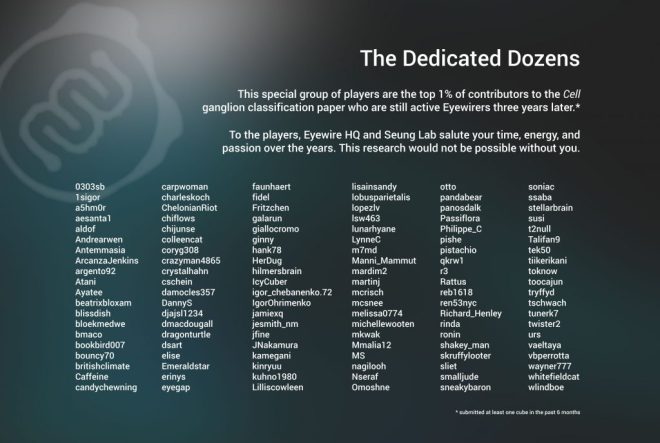
The top 10% of players (2,930 people) contributed 87% of the total cube submissions. The top 1% (293 people) contributed a whopping 52% of the total cube submissions.

Among the top 10% of players, 15% still play Eyewire after three years. We love you guys!

For Science!
The new cells are so new that not much is known about what they do. Here’s a summary of what we know so far.
Ganglion cells are divided up mostly based on where within the retina their branches can be found. Specifically, all ganglion cells have branches in the inner plexiform layer aka the IPL.
The ganglions are divided up into 47 clusters, which you can see in this graphic:

The names may sound weird, but with so many cells identified in part by their dendritic stratification profile (where their branches are within the retina), an anatomically-based naming schema comes in handy. You can use this key to figure out just what names like 7ir and 37c mean:

Outer — Inner (o,i): Relative location of peak dendritic density within the retinal Inner Plexiform Layer (IPL)
Depth (1-9): Specific depth within the IPL. 1 is closest to Ganglion Cell Layer & 9 is closest to the Inner Nuclear Layer
Short — Tall (s,t): Relative height span of dendritic arbor.
Narrow — Wide (n,w): Relative width span of dendritic arbor.
Asymmetry (a): Indicates asymmetry in dendritic branches
Dorsal, Caudal, Ventral, Rostral: Cardinal neural directions
The ganglion cells are divided up into six broad categories based generally on the location of the majority of their dendrites within the inner plexiform layer of the retina.

Knowing where the branches stratify gives us clues about their functional attributes, such as what visual stimuli causes the cells to fire. The lab will use this information to dive deeper into the new cell types and to research more about the new ganglion cell classification contained in this publication.
Let us know if you have any questions and thanks again for being a part of the Eyewire community. If you’re ready to jump into the next chapter, be sure to sign up to be a beta tester for Neo, a new brain mapping game that will come out later this year.
For science!

