New Eyewire Publication in Cell: A Classification for Retinal Ganglion Cells
A New Classification of Ganglion Neurons

An exciting new prepublication paper from the Seung Lab squad is available on biorxiv! We’ve been working for several years to map and analyze neurons from e2198, the dataset of Eyewire. Along the way…
A new metric
How many types of cells are there? Hard to answer if you don’t even have a definition for what makes a cell type. Researchers argue over what makes a cell — its shape? Activity? Connections? Perhaps even a combination. A greater question may be: how do we empirically say “this cell is different from that cell,” when so many neurons look almost the same?
We coalesce multidimensional data about the neurons, such as where they branch (stratification profile), when they fire (temporal response) and what stimuli makes each cell class fire (direction selectivity), in order to get the big picture of these tiny cells. This paper applies Density Conservation, a metric that averages dendrite branches within a given volume, to clusters of cells in order to guide classification. We divide our dense sample of ganglion cells into six high-level clusters, which are subdivided further to end up with 47 clusters, all of which you can explore on Museum.
What does it all mean?
If this method becomes more broadly accepted, then so too will the types of cells it has helped identify. Six new cell types, to be exact! The types currently bear the following lackluster names: 1ni, 1no, 2o, 85, 27, 5to, explained in the key below. And here they all are in interactive 3D on the museum.
Neurons named by Eyewirers
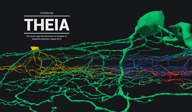
Type 27 is named Theia
Museum Tour Guide
Cells in the museum have a naming structure that’s convenient for researchers but they aren’t so easy to understand at first glance, so we created this key and the following descriptions that explain the names and graphs.

Short/Tall: Height of dendritic arbor — a neuron’s branches
Depth: location within the IPL, the Inner Plexiform Layer, of the retina, where the Eyewire dataset comes from
Outer/Inner: Relative location of dendritic arbor (note this is not inner/outer layer of IPL)
Wide/Narrow: dendritic arbor width
Dorsal/Ventral/Caudal/Rostral refer to the direction preference of the neurons themselves.
—
The trickiest chart to understand in Museum is Directional Response. Cells in the retina respond to things an animal sees — incoming stimuli is derived from photons bouncing off an object that can literally be moving. Think about all the ways things you see can move. People walk left and right; an airplane flies by overhead while a sparrow lands below. When it comes to the retina, these stimuli are not treated equally. Specific cells respond only to motion detected in specific directions.

The four charts at the left together show neurons that will respond to any direction of movement; however, each type of cell 37 responds to a discrete and unique direction of movement, shown in type 37c by an arrow. The chart at right shows cell type 4on. It has no preferred direction, so we say it is not a directionally selective cell. Note that the direction preference is opposite (37r prefers caudal direction) because the lens flips what the eyes see. Explore these five cells in the museum.
Stratification Profile
Depth within the IPL where a cell’s branches are found.

The IPL is subdivided into four sublaminae, the location of each indicating aspects of the cells whose dendrites arborize in that region. The On layer contains dendrites that respond when a new bit of light appears in an animal’s visual field. The Off layer houses just the opposite: cells that fire when a bit of light leaves field of view. Some cells are bistratified, with branches across both layers, and they respond when light appears and disappears. Many cells striate in just a single layer. See these specific cells in the museum.
You can see how cells fire with respect to light be checking out the Temporal Response chart in Museum.

Temporal response is also a bit tricky to understand. Think of a dark night when you look up at the sky and a shooting star suddenly streaks by. Cells in the left chart that fire at time 1 for ON would spike when your eyes detect the presence of this shooting star that wasn’t there before. When do you think cells from the right chart would fire? They respond to OFF stimuli – visual stimulus that was there but is no longer – so they would fire after the shooting star has disappeared. The axons of these cells converge to form the optic nerve then dive deep into the brain. Certain cells tell your brain something new has appeared while others say something you were seeing is gone. And there are cells that respond to both — see if you can find them in the museum!
The cells studied were mapped by Eyewirers during the Neuropia Countdown.
Food for thought: these 47 types of cells are all found in the retina..researchers think there may be hundreds or thousands of types of neurons in the rest of the central and peripheral nervous system!
What’s next? Go exploring in the museum and gain a better understanding of the science you’ve made possible!
Click the images below to jump to those cells in museum.

